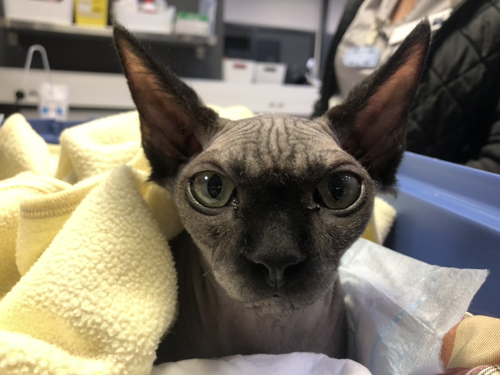
Toscane, the Sphynx cat who underwent a faecal transplantation
Toscane, a 9mth old female neutered Sphynx cat
Before visiting the Specialist.
Animal Treated
Cat
Animal Condition
Mass in mouth
Specialist(s) Required
Veterinary Pathology
The work of the Specialist behind the scenes, in this case a Pathology Specialist, is highlighted in the case of Tigger.
Before visiting the Specialist
Tigger is a 14 year old, male (neutered) Domestic short haired cat, who was taken to his primary vet because he was having difficulty eating his food. When the vet examined Tigger’s mouth he noticed Tigger had signs of dental disease affecting some of his teeth and more worrying than that, there was an angry-looking red mass on the gums of his bottom jaw. The vet decided that Tigger needed a general anaesthetic in order to investigate this mass further.
Tigger’s Care
Under general anaesthesia, the vet examined Tigger and took a X-ray, which showed that the mass was destroying some of the underlying bone of the jaw. It was vitally important for Tigger’s ongoing care to find what type of mass was affecting Tigger’s jaw. Was it malignant or benign? Was it likely to spread to other parts of Tigger’s body? The vet took a biopsy from the mass and submitted it to a diagnostic laboratory, where a Specialist in Veterinary pathology did the microscopic assessment of the mass. Under the microscope the Pathology Specialist saw lots of bizarre-looking giant cells which contained multiple nuclei (called multinucleate giant cells), as well as more spindle-shaped cells which were obviously dividing. These cells were in amongst lots of fibrous tissue which in places was beginning to turn into bone. With this combination of features, together with the location within the mouth and Tigger’s history, a diagnosis could be made of a “peripheral giant cell granuloma” or a “giant cell epulis”
Tigger’s Progress
For Tigger, this is quite a good diagnosis – although it is possible the mass might grow back over time, and the vet may have to surgically remove it again, it is generally not considered to be malignant. These masses can develop at any age in humans and the same is thought to be true in animals; we still don’t know the precise cause, but it might be a response of the tissues to the presence of foreign material or tooth disease. This case is a very good example where close collaboration between the veterinary surgeon and the Veterinary Pathology Specialist enables the vet to give Tigger the best possible care.
Article provided by Melanie Dobromylskyj
Specialist in Veterinary Pathology
www.finnpathologists.co.uk